Blog
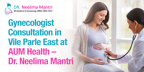
Gynecologist Consultation in Vile Parle East at AUM Health – Dr. Neelima Mantri
Women seeking reliable and compassionate gynecological care in Vile Parle East, Mumbai, can consult Dr. Neelima Mantri at AUM Health. The clinic is known for providing personalized women’s healthcare with a focus on comfort, clarity, and clinical excellence. About Dr....
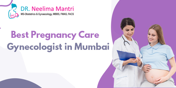
Best Pregnancy Care Gynaecologist in Mumbai
Pregnancy brings joy mixed with worry when you want everything to go perfectly for nine months until your baby arrives safely. Choosing the right doctor matters tremendously because pregnancy care shapes outcomes for both mother and child through every stage. The best...
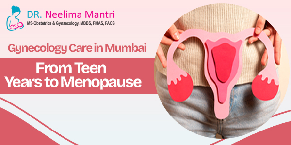
Gynecology Care in Mumbai From Teen Years to Menopause
Women go through major body changes from their first period all the way through menopause decades later in life. Each phase brings different health questions and concerns that need a doctor who actually gets what you are dealing with right now. Finding the Best...
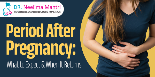
Period After Pregnancy: What to Expect and When It Returns
Pregnancy changes the body in many ways. One of the most common questions women ask after giving birth is when their period will return. Some expect it to come back within weeks, while others may not see it for several months. Every experience is different. That is...
Early Warning Signs of PIH Every Pregnant Woman Should Know
Pregnancy is a significant development in every couple's life. It brings joy along with unexpected complications. High blood pressure is one such complication that begins after the fifth month. This condition is called Pregnancy-Induced Hypertension [or PIH]. It often...
Is Norethisterone Safe for Period Delay? Understanding Its VTE Risk
Delaying your period can feel like a simple choice when you are planning a trip, wedding, or exam schedule. Many women turn to medication for this purpose, especially if their cycle is regular but inconvenient. One such option is norethisterone. It is often prescribed...
How Age Affects Ovulation and Fertility in Women
Fertility is shaped by many factors, but a woman’s age remains one of the most important. As the years pass, subtle changes begin to affect ovulation and reproductive capacity. These changes are biological and cannot be paused. Many women learn about them only after...
When Should You Get a Follicular Scan? Best Timing for Fertility Monitoring
A follicular scan helps track ovulation in real time so you can plan your pregnancy with greater accuracy. Many women often ask, “When should I get a follicular scan?” Because timing decides how useful the scan will be for fertility planning. The process maps egg...
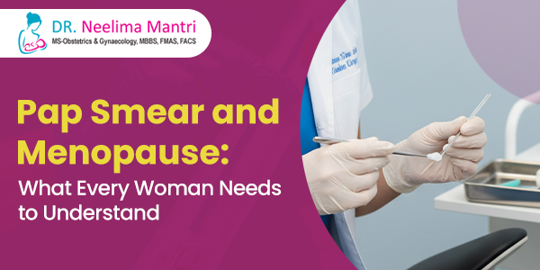
Pap Smear and Menopause: What Every Woman Needs to Understand
Many women assume that routine tests like a Pap smear are no longer required once menopause begins. The truth is that menopause introduces hormonal changes that can silently impact cervical health. Skipping screenings during this stage can allow risks to go...
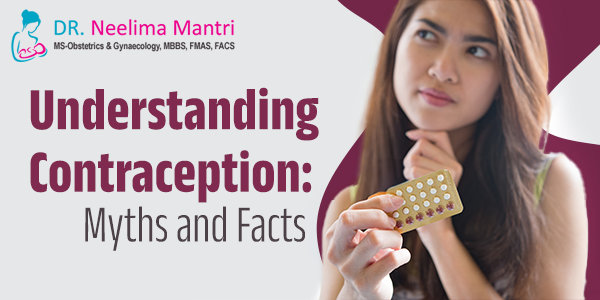
Understanding Contraception: Myths and Facts
Contraception is one of the most common health topics discussed during gynaecology visits. Still, it remains surrounded by confusion and half-truths. Some people avoid it due to fear. Others follow advice passed from friend to friend without medical checks. This makes...
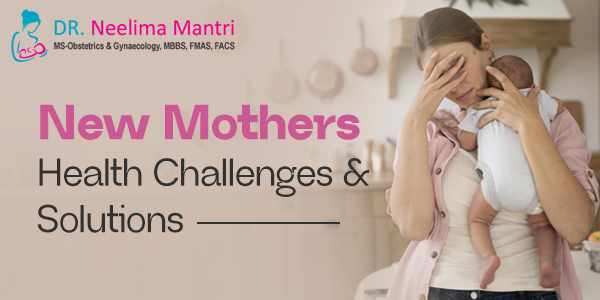
New Mothers’ Health Challenges and Solutions
The journey of childbirth brings both joy and stress. While the baby becomes the focus of everyone around, the mother's body goes through silent changes that are often overlooked. These changes are not just physical but emotional as well. From body aches to mood...
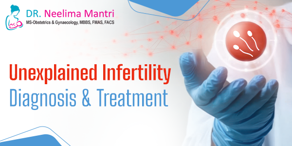
Unexplained Infertility Diagnosis and Treatment
You are trying to conceive. You have done all the right things. The tests seem normal. But still, there is no pregnancy. This is often where the term “unexplained infertility” comes in. It means everything looks fine on paper, yet conception does not happen. It can...